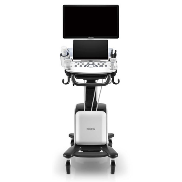
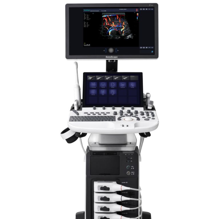
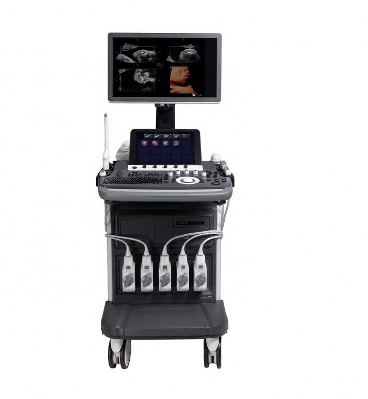
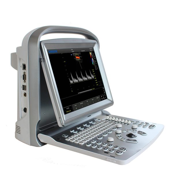
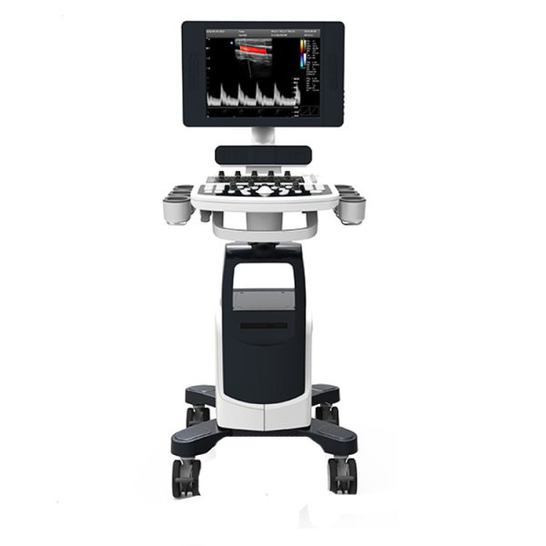
Ultrasonido abdominal chison i8 : Ultrasonido 4d doppler obstetrico chison i8 Sistema Doppler Color Digital Ficha técnica V1.0 28 de marzo de 2014 Ultrasonido abdominal chison i8 Dimensiones y peso * Dimensiones de la unidad principal (aprox.): 825 mm x 519 mm x 1458 mm * Peso neto de la unidad principal (aprox.): 90 kg (sin sonda incluida) Alimentación eléctrica * Voltaje de la fuente de alimentación: Adaptable automáticamente para CA 100 V-240 V * Frecuencia de la fuente de alimentación: 50-60 Hz * Consumo de energía: 600 VA Información general Ultrasonido abdominal chison i8 Panel de operación * Panel de control * Teclado alfanumérico * 8 diapositivas TGC * Teclas retroiluminadas interactivas * LCD a color de alta resolución – Dimensión diagonal: 19 pulgadas – Resolución: 1280X1024 – Ajuste de brillo * Altavoz integrado – Volumen ajustable Interfaz de usuario Ultrasonido abdominal chison i8 Aplicaciones * Abdomen * Ginecología * Obstetricia * Urología * Partes pequeñas * Pediatría * Vascular * Musculoesquelético * Método de escaneo cardíaco * Convexo electrónico * Lineal electrónico * Microconvexo electrónico * Matriz en fase electrónica * Convexo volumétrico Tipos de transductores * Transductor convexo de 3,5 MHz: D3C60L * Transductor lineal de 7,5 MHz: D7L40L * Transductor lineal de 7,5 MHz: D7L60L * Transductor lineal de 12,0 MHz: D12L40L * Transductor transvaginal de 6,0 MHz: D6C12L * Transductor transvaginal de 7,5 MHz: D7C10L * Transductor microconvexo de 3,0 MHz: D3C20L * Transductor microconvexo de 5,0 MHz: D5C20L Descripción general del sistema * Transductor microconvexo de 6,0 MHz: D6C15L * Transductor de matriz en fase de 3,0 MHz: D3P64L * Transductor de matriz en fase de 6,0 Mhz: D6P64L * Transductor de volumen 4D de 4,5 MHz: V4C40L Modos de imagen * Modo B * Modo 2B * Modo 4B * Modo B/M * Modo M * Modo THI * Modo B/BC * Modo CFM (Mapeo de flujo de color) * Doppler de pulso * CPA (Doppler de potencia) * DPD (Doppler de potencia direccional) * Imágenes compuestas múltiples * SRA (Algoritmo de reducción de moteado) * i-Imagen * Imágenes trapezoidales (solo para transductor lineal) * Imágenes panorámicas curvas * Dirección 2D * Superaguja * Elastografía * Triplex * Trazo y medición PW automáticos en tiempo real * Cuádruple * Croma B&M&PW * CWD * Modo M color * TDI * Modo M de dirección libre * IMT * ECG * 4D * Modo de visualización DICOM * Pantalla cuádruple/dual (para B, CFM, CPA) * Modo dúplex: B+CFM, B+PW, B+CPA,B+DPD,B/M * Modo triplex: B+CFM+PW, B+CPA+PW,B+DPD+PW, B+CFM+CW, B+CPA+CW, B+DPD+CW * Modo cuádruple: B+C+PW+ Trazo y medición de PW automáticos en tiempo real Anotación en pantalla * Nombre del hospital * Fecha/hora * Nombre e ID del paciente * Estado del sistema (en tiempo real o congelado) * Barra de grises/color * Guía de cine * Dirección de escaneo * Ventana de resumen de medición * Ventana de resultados de medición * Tipo de transductor * Frecuencia * Nombre de la aplicación * Indicación de menú * Indicación de funciones del trackball * Parámetros de imagen mostrados en la pantalla * TDI * Configuración estándar de CW * Pantalla LCD de 19 pulgadas de alta resolución * 4 puertos de transductor activos * Imágenes de flujo Doppler color * Doppler de onda de pulso * Imágenes de flujo Doppler de potencia * Imágenes de flujo Doppler de potencia direccional * Disco duro integrado ≥250G * Puertos USB: 6 (2 en el panel de control, 4 en el panel trasero) * Puerto Ethernet * Puerto de salida S-video * Puerto VGA * Interruptor de pedal * Puerto de salida de audio * Control remoto * Puerto de salida de video * Paquete de medición general * Paquete de medición clínica * Pantalla en varios idiomas * EASYVIEW TM: sistema de archivo de imágenes * Sistema de gestión de información del paciente * Sistema de informes de edificios * AIO (Optimización automática de imágenes) * Zoom inteligente * Algoritmo de reducción de moteado (SRA) * Paquete de software i-Image TM * Trazo de espectro Doppler automático y cálculo automático Opciones de software * DICOM 3.0: Almacenamiento, Impresión, Lista de trabajo, MPPS, Almacenamiento SR * Paquete de software 4D * Elastografía * Imágenes panorámicas curvas * Superaguja * Paquete cardíaco: CW, Modo M de dirección libre, Modo M de color, TDI, ECG * IMT * Opción de hardware 2D steer ultrasonido abdominal chison i8 * Transductor convexo de 3,5 MHz: D3C60L * Transductor lineal de 7,5 MHz: D7L40L * Transductor lineal de 7,5 MHz: D7L60L * Transductor lineal de 12,0 MHz: D12L40L * Transductor transvaginal de 6,0 MHz: D6C12L * Transductor transvaginal de 7,5 MHz: D7C10L * Transductor microconvexo de 3,0 MHz: D3C20L * Transductor microconvexo de 5,0 MHz: D5C20L * Transductor microconvexo de 6,0 MHz: D6C15L * Transductor de matriz en fase de 3,0 MHz: D3P64L * Transductor de matriz en fase de 6,0 MHz: D6P64L * Transductor de volumen 4D de 4,5 MHz: V4C40L * Módulo CW * Módulo 4D * Kit de biopsia: para sonda convexa/lineal/TV respectivamente * Periféricos de pedal * Impresora de vídeo: SONY UP-897MD,SONY UP-D25MD * PC impresora: – HP Laser Jet 1020 – HP Laser Jet CP2055d Modo B * Potencia acústica * Ganancia * TGC * Profundidad * Frecuencia * Velocidad de fotogramas * Número de enfoque * Posición de enfoque * Ancho de escaneo * Densidad * Dinámico * Persistencia * Rechazo de ruido * Suavizado * Mejora de bordes * i-Image TM * SRA * Compuesto * Mapa 2D * Croma * Gamma * Brillo de pantalla * Rotación de imagen * Voltear (izquierda/derecha, arriba/abajo) * Zoom * Dirección 2D * Procesamiento y presentación de imágenes de elastografía * Superaguja * Imágenes trapezoidales (solo para transductor lineal) Modo M * Mapa de color * Velocidad de barrido * Diseño * Dirección libre Modo M Modo de color * Ganancia * Frecuencia * Velocidad de fotogramas * Dirección * PRF * Filtro de pared * Mapa de color * Velocidad de flujo * Inversión de color * Densidad * Persistencia * Línea base * Modo de color: Velocidad, Varianza * Efecto de sangre * Escala * Tamaño de paquete * Thre de pared Modo CPA/DPD * Ganancia * Frecuencia * Velocidad de cuadros * Dirección * PRF * Filtro de pared * Mapa de color * Flujo * Densidad * Persistencia * Thre. de pared * Tamaño de paquete Modo PW * Ganancia * PRF * Escala * Invertir * Filtro de pared * Audio * Velocidad * Línea base * DA * SV * Mapa de color * Mapa 2D * Mejora de espectro * Rango dinámico * Triplex * DV media * DV máx. * Auto Cal * D Trace Smooth * Umbral * Área de traza Modo CW * Ganancia * PRF * Escala * Invertir * Filtro de pared * Audio * Mapa de color * Velocidad * Línea base * Mapa 2D * CWD FFT * Mejora CWD * Dinámico * DA Cineloop * Soporte 2D, M, PW, CFM, CPA, DPD, CW, Color M, Dirección libre M * Revisión simultánea e independiente en modo dúplex * Cineloop automático/manual * Velocidad de reproducción de cine variable * Marco de inicio y final de almacenamiento de cine definido por el usuario * Marco de inicio y final de revisión de cine definido por el usuario * Almacenamiento permanente en disco duro y visualización en modos en tiempo real * Presentación de diapositivas: función de presentación de diapositivas Almacenamiento * ≥250 GB de disco duro integrado * Controlador de DVD R/W * Puertos USB * Formato de almacenamiento de imágenes fijas: IMAG * Formato de exportación de imágenes fijas: BMP, JPG, DCM, PNG, TIFF * Formato de almacenamiento de bucles de cine: CINE * Formato de exportación de bucles de cine: AVI * Configuración de almacenamiento rápido: 3 s, 5 s, 10 s, tiempo personalizado, manual EASYVIEW TM * Revisión de imagen Diseño: 1 × 1, 2 × 2 * Gestión de imágenes – Eliminar imagen seleccionada – Exportar imagen seleccionada – Enviar imagen seleccionada a demostración – Imprimir imagen seleccionada por impresora de PC – Imprimir imagen seleccionada por impresora DICOM – Enviar imagen seleccionada por DICOM – Seleccionar todo – Seleccionar ninguno Revisión de examen * Buscar examen * Revisión de examen: vista del paciente, vista del estudio * Gestión de exámenes – Eliminar examen seleccionado – Exportar examen seleccionado – Hacer copia de seguridad del examen seleccionado – Recuperar del examen de copia de seguridad – Seleccionar todo – Expandir todo – Contraer todo – Editar examen seleccionado – Revisar examen seleccionado – Continuar examen seleccionado Paquete de medición general de ultrasonido abdominal chison i8 – Paquetes de software para varios usos clínicos específicos – Métodos de análisis integrales – Informes de análisis clínicos * Paquete de medición general * Medición normal en modo B Distancia Longitud_Área(Elipse) Longitud_Área(Trazo) Volumen(1 Distancia) Volumen(2 Distancia) Volumen(3 Distancia) Volumen(1 Elipse) Volumen(2 Elipse) Volumen(1 Distancia 1 Elipse) Relación Ángulo * Medición normal en modo M Distancia M Tiempo M Velocidad Frecuencia cardíaca * Medición normal en modo PW Velocidad Distancia Pico Trazo automático Trazo manual Medición y cálculo de FC Flujo Volumen StD% StA% Área ICA/CCA Paquetes de análisis clínicos * OB Medición OB -B Distancia Biometría fetal: GS, CRL, YS, BPD, OFD, HC_Ellipse, APD, TAD, AC(Elipse), FTA, FL, SL, APTD, TTD, ThC Huesos largos fetales: Húmero, ULNA, Tibia, RAD, FIB, CLAV Cráneo fetal:CER, CM, NF, NT, OOD, IOD, NB, LVent, HW OB Otros: Lt Kid, Rt Kid, Lt Renal AP, Rt Renal AP, LV Wr HEM, MAD AFI: AFI_1,AFI_2,AFI_3, AFI_4 FBP: AF Ductus venoso: StA%, StD%, Área del vaso, Dis del vaso StA%: A Fuera, A En StD%: D Fuera, D En CX_L Aorta: StA%, StD%, Veslumen_D,Veslntimal_D,VesOutside_D,Veslntimal_A, Veslumen_A StA%: A Fuera, A En StD%: D Fuera, D En Aorta descendente: StA%, StD%,Veslumen_D,Veslntimal_D,VesOutside_D,Veslntimal_A ,Veslumen_A StA%: A Out, A In StD%: D Out, D In MCA: StA%, StD%,Veslumen_D,Veslntimal_D,VesOutside_D,Veslntimal_A,Veslumen_A StA%: A Out, A In StD%: D Out, D In Umb A: StA%, StD%, Veslumen_D,Veslntimal_D,VesOutside_D,Veslntimal_A, Veslumen_A StA%:A Out, A In StD%: D Out, D In Arteria uterina: Arteria uterina (Rt), Arteria uterina (Lt) Arteria uterina (Rt): StA%, % estándar, Veslumen_D, Veslntimal_D, VesOutside_D, Veslntimal_A, Veslumen_A Arteria uterina (Izq.): StA%, StD%, Veslumen_D, Veslntimal_D, VesOutside_D, Veslntimal_A, Veslumen_A StA%: A Salida, A Entrada StD%: D Salida, D Entrada Arteria pulmonar: StA%, StD%, Veslumen_DV, Veslntimal_D, VesOutside_D, Veslntimal_A, Veslumen_A StA%: A Salida, A Entrada StD%: D Salida, D Entrada Selección fetal Medida OB -D Umb A Aorta Aorta descendente Arteria uterina (Izq.) Arteria uterina (Der.) Arteria pulmonar ACM FCF Medida OB –M MDistancia MTiempo Velocidad Frecuencia cardíaca * GYN GYN -B mide Distancia UT: UT_L, CX_L, UT_W, UT_H Vol. Cervix: Longitud, Altura, Ancho ENDO Volumen OV_Derecho: Longitud, Altura, Ancho Volumen OV_Izquierdo: Longitud, Altura, Ancho FO_D Derecho: Longitud, Ancho FO_D Izquierdo: Longitud, Ancho Arteria uterina: Arteria uterina(Der), Arteria uterina(Izquierda), Arteria uterina(Der): StA%, StD%, Área del vaso, Disposición del vaso Arteria uterina(Izquierda): StA%, StD%, Área del vaso, Disposición del vaso StA%: A Salida, A Entrada StD%: D Salida, D Entrada GYN -D mide Umb A: Umb A(Der), Umb A(Izquierda) MCA: ACM (Dt), ACM (Izq.) A uterina dcha.: A uterina dcha. (Dt, A uterina dcha. (Izq.) A uterina izq.: A uterina izq. (Dt, A uterina izq. (Izq.) AO fetal: AO fetal (Dt), AO fetal (Izq.) FCF Medida GINECÓMICA-M Distancia M Tiempo Velocidad Frecuencia cardíaca * Pediatría HIP * URO Distancia Vol residual Vol. prostático Riñón izquierdo Riñón derecho Vol. zona T Vol. vejiga Est. A% Dest. Est. % Área vascular Dis. vascular * Cardíaco Medida Cardíaca-B Distancia Plano único Biplano Volumen bala Modificación_Simpson Cubo de Teichholz VI/VD AO/VI TSVI MV AV Medida Cardíaca-D TSVI MV AV MV TV PV Vena pulmonar FC Medida Cardíaca-M Distancia Frecuencia_Corazón Tiempo_Eyección LV LVSHORT AV AVSHORT MV AV AO/LV LVOT TV PulV * Vaso Auto IMT Prox CCA CCA Media CCA Distal ACI Prox ACI Media ACI Distal ECA A Vertebral A INT IIL EXT IL ILIAC CFA ProFun LTCIR SFA Pop A ATA PTA PERON DRPED * Abdomen CBD GB Pared Hígado Longitud Aorta Prox Aorta Media Aorta Distal Bazo Vol. Renal Lliac * Carótida A Subclavia A Prox CCA CCA Media CCA Distal Bulbo ACI Prox ACI Media ACI Distal ECA A Vertebral A Medición General Flujo Volumen * Piezas Pequeñas Medición General Relación Ángulo Mediante la configuración del sistema, los usuarios pueden * Personalizar la información del hospital * Personalizar el idioma * Personalizar el tiempo de almacenamiento rápido * Personalizar el mapa de colores * Asignar funciones al botón “IMPRIMIR” en el panel de control y el pedal * Personalizar la biblioteca de comentarios * Personalizar el informe Funciones definidas por el usuario Por definición del usuario función, los usuarios pueden personalizar el ajuste predefinido por el usuario, incluyendo: – Nombre de las aplicaciones, Nombre de los ajustes predefinidos, Nombre definido por el usuario – Tipo de examen de las aplicaciones – Parámetros de imágenes Interfaz de pantalla en varios idiomas * Inglés * Chino * Polaco * Portugués * Ruso * Español * Danés * Alemán * Francés Sistema operativo Windows XP Embedded Configuración del sistema * Entrada de alimentación de CA: 1 * Salida de audio: L、R * S-video: 1 * Salida de video: 1 * VGA: 2 * Puerto USB: 6 * Ethernet: 1 * Control remoto: 1 * Puerto del pedal: 2 * Salida de CA: 1 * Poste de tierra: 1 * Botón de encendido: 1 Entradas y salidas * Temperatura ambiente: 10 °C a 40 °C * Humedad relativa: 30 % a 75 % * Presión atmosférica: 700 hPa a 1060 hPa Condiciones de funcionamiento * Temperatura ambiente: -5 °C a 40 °C * Humedad relativa: ≤ 80 % (sin condensación) * Presión atmosférica: De 700 hPa a 1060 hPa. Es posible que no todas las características o especificaciones descritas en este documento estén disponibles en todas las sondas o modos. CHISON Medical Imaging Co., Ltd. se reserva el derecho de modificar las especificaciones y características que se muestran aquí, o de discontinuar el producto en cualquier momento sin previo aviso ni obligación. Para obtener la información más actualizada, comuníquese con un representante de CHISON. Condiciones de almacenamiento. Oferta especial de ultrasonido Sonoscape | Lista de precios de ultrasonido Chison. Precio de SonoScape X5. Precio de SonoScape S9. Precio de Sonoscape S8. Precio de Sonoscape S8 EXP. Sonoscape A5. Sonoscape S2. Sonoscape A6. Sonoscape S6. Sonoscape A6VET. Sonoscape S40. Sonoscape S50. Sonoscape S22. Sonoscape S12. Sonoscape S30. Sonoscape S11. Precio de SonoScape X3. Precio de Sonoscape S20. Precio de Chison Q5. Precio de Chison Q9. Precio de Chison Ebit 50. Precio de Chison Ebit 60. Precio de Chison I3. Chison I8. Precio Chison EC01, precio Chison EC03, precio Chison EC05, precio Chison EC06, imagen del equipo B.S.D., certificado B.S.D., B.S.D. Medical colabora con DHL, FEDEX, UPS, EMS, TNT, etc., empresas de transporte internacionales que garantizan la llegada segura y rápida de sus productos a su destino.

 Retiro en Casa Matriz
 Envío a Domicilio
 Certificado de Calidad
 Empaque y Traslado Seguro