
-
 Retiro en Casa Matriz
-
 Envío a Domicilio
-
 Certificado de Calidad
-
 Empaque y Traslado Seguro
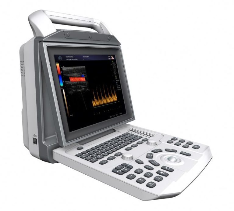
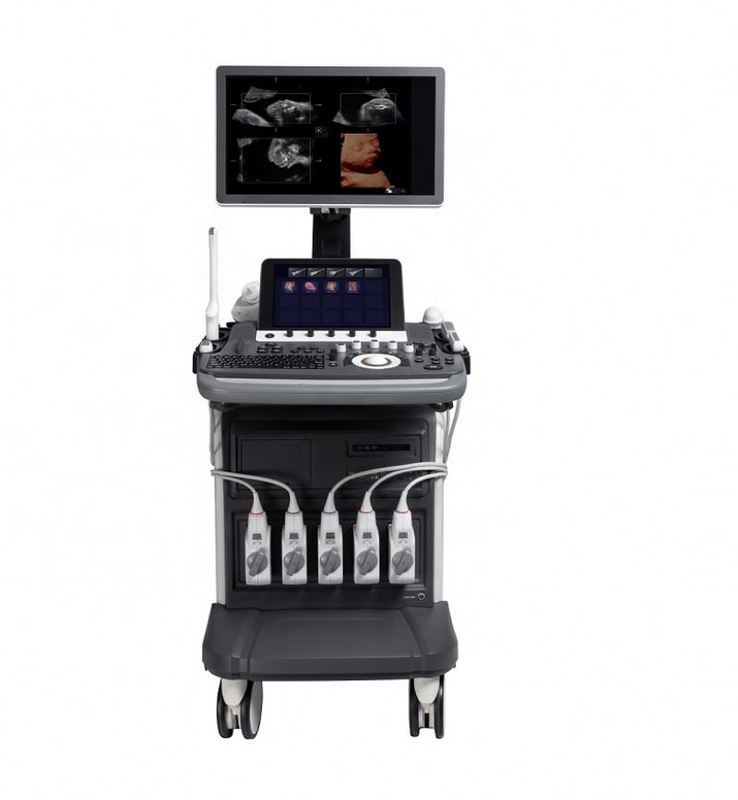
Doppler vascular SonoScape S40 Configuraciones estándar El hardware estándar incluye: Unidad principal S40 Monitor LED a color de alta resolución de 19″ Pantalla táctil de alta resolución de 10″ Soporte de sonda endocavitaria Cinco conectores de transductor Un conector de transductor CW DVD-RW/USB 2.0/Disco duro Módulo de ECG de 500 G El software estándar incluye: Modos de imagen: B/2B/4B/M/THI/CFM/PDI/DirPDI/PW/HPRF/CW LGC: Compensación de ganancia lateral Tecnología multihaz Inversión de pulso μ-Scan armónico: Tecnología de reducción de moteado 2D Imágenes compuestas Imágenes trapezoidales Imágenes panorámicas 2D Imágenes panorámicas en color Imágenes 3D a mano alzada S-Depth 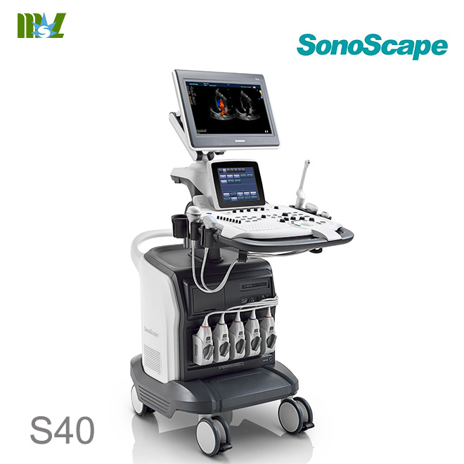 Kit cardiovascular avanzado S-Live Auto NT: TDI/ Color M/ IMT/ Steer M/ Ecocardiograma de estrés automático VIS-Needle M-Tuning: optimización de imagen con un solo botón DICOM 3.0: Store/C-Store/Worklist/MPPS/Print/Q/R Transductores configurados estándar: Matriz convexa de 128 elementos 3C-A (abdominal, obstetricia, ginecología), 1.0-7.0 MHz/ R50 mm Matriz lineal de 192 elementos L742 (vascular, partes pequeñas, MSK, etc.), 3.5-16 MHz/ 38 mm Matriz en fase de 80 elementos 4P-A (cardíaco, transcraneal), 1.0-6 MHz Matriz endovaginal de 192 elementos 6V3 (ginecología, obstetricia, urología), 3-15 MHz/ R10 mm
 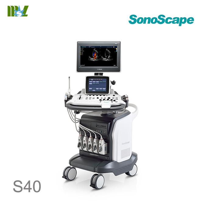 Doppler vascular SonoScape S40 video Doppler vascular SonoScape S40 Configuraciones opcionales Estático 3D 4D Imágenes de contraste C-xlasto: Imágenes de elastografía Doppler vascular SonoScape S40 Especificaciones generales El sistema de ultrasonido Doppler color S40 adopta las tecnologías Doppler ultrasónicas avanzadas, que incluyen el formador de haz de banda súper ancha completamente digital, enfoque dinámico digital, apertura variable y rastreo dinámico, rango dinámico de banda ancha, procesamiento paralelo de múltiples haces, etc. El sistema de software de diagnóstico por ultrasonido, las imágenes del sistema de ultrasonido, las interfaces de operación en varios idiomas y la pantalla táctil con tecnología de interacción persona-computadora se pueden personalizar fácilmente de acuerdo con el diseño de la ingeniería humana. Los usuarios pueden utilizar el sistema con un mínimo de formación o guía. Este sistema ha sido diseñado para cumplir con las normas y regulaciones internacionales aplicables, garantizando así la seguridad y disponibilidad del producto. Se basa en tecnología informática y el sistema operativo Linux, lo que lo hace más flexible y estable. El mantenimiento y la actualización de funciones del sistema se pueden realizar mediante actualizaciones de software, lo que aumenta el valor del producto y mantiene el avance tecnológico. Tecnologías avanzadas del sistema de ultrasonido Doppler color SonoScape S40 *Tecnología digital de vanguardia *Tecnología de procesamiento multihaz *Imágenes compuestas espaciales *Tecnología de posprocesamiento *Imágenes armónicas de tejidos *Alta frecuencia de repetición de pulsos *Imágenes panorámicas *Imágenes 4D *Icono gráfico de diagnóstico *Pantalla táctil con tecnología de interacción persona-computadora *Sistema de elevación del teclado Configuraciones estándar del sistema de ultrasonido Doppler color SonoScape S40 *Modo B *Modo color *Modo PW *Modo CW *Modo THI *Modo TDI *Modo DPI *Modo DDPI *Imágenes 3D *Paquete de medición de cardiología *Paquete de medición de ginecología *Paquete de medición de urología *Paquete de medición vascular *Paquete de medición de piezas pequeñas *Paquete de medición ortopédica *Medición IMT *Índice TEI *Trazado automático Doppler espectral *Medición del volumen de flujo de color *Sonda MLA *Sonda de matriz en fase Especificaciones del sistema de ultrasonido Doppler color S40 2 *Tecnología de procesamiento multihaz *Función μ-scan *Función TDI *Modo B: cinco frecuencias variables *Alta frecuencia de repetición de pulsos *Modo triplex *Imágenes panorámicas *Imágenes compuestas *Imágenes trapezoidales *Grabadora de DVD RW *Módulo de función de ECG 4. Funciones opcionales *Modo Steer M *Modo Color M *Imágenes 4D *Sonda TEE *Función de rotación de imagen *Medición de IMT *Función B Flow *Transmisión DICOM *Compromiso de almacenamiento DICOM *Función de lista de trabajo DICOM *MPPS *Eco de esfuerzo 5. Accesorios opcionales *Soporte de biopsia *Impresora de inyección de tinta a color *Impresora de video B/N *Impresora de video a color *Colgador de cable de sonda 6. Rangos de escaneo de la sonda *Transductor curvo: ≥70° *Transductor de matriz en fase: ≥90° *Transductor microcurvo: ≥193° 7. Métodos de escaneo *Escaneo electrónico de sector curvo *Escaneo electrónico de matriz lineal *Escaneo electrónico de sector de matriz en fase 8. Aplicaciones *Abdomen *Vascular *Cardiología *Ginecológico/Obstétrico *Urología *Musculoesquelético *Ecografía intervencionista *Partes pequeñas *Anestesiología 9. Modos de imagen *Modo B *Modo M *Modo THI *Modo CDI *Modo DPI *Modo TDI *Modo PW Especificaciones para el sistema de ultrasonido Doppler color S40 3 *Modo CW *Modo 3D/4D *Modo Color M *Modo Steer M 10. Formatos de visualización *Dual B *Quad B *B + PW *B + CW *B + M *B + Color *Dual B (Flujo) *B + Color + PW *B + Color + CW *B + Color M *Imágenes panorámicas *Imágenes trapezoidales 11. Menú de configuración del sistema *Historial de exámenes *Nuevo examen *Continuar *Revisar Seleccionar todo Almacenar en DICOM Almacenar en USB Eliminar Imprimir Informe Salir *Eliminar *Almacenar *Imprimir *Confirmar *Salir *Ajustes del sistema *Ajustes generales Nombre de la instalación Idioma Inglés Chino simplificado Español Ruso Francés Italiano Alemán Turco Protector de pantalla Sensible al trackball Formato de clip MP4, AVI Formato de imagen JPG, BMP, TIF Protector de pantalla: Ajustable Color de ROI Verde, Amarillo Naranja, Azul Formato de visualización: H1/2, H1/4, V1/3, V1/2, V2/3, O1/4 Guardar con una tecla: Activado/Desactivado Especificaciones para el sistema de ultrasonido Doppler color S40 4 Unidad EFW: Seleccionable Formato de fecha mm/dd/aaaa aaaa/mm/dd dd/mm/aaaa Formato del informe PDF, TXT Número de fotograma guardado: Ajustable *Establecer impresora Controlador de impresora Invertir vídeo Insertar controlador *Establecer menú de cálculo Modo 2D Ángulo Volumen Volumen L×An×Al Área Doppler Flujo de color IMT Vascular Parte pequeña Ortopédico Obstétrico/Ginecológico Ventrículo izquierdo Urológico Evaluación de la salud de la arteria carótida Evaluación de la salud de la arteria carótida (CF) Diámetro de la válvula mitral Diámetro de salida del VI Diámetro de la válvula pulmonar Diámetro de la aorta Modo PW Velocidad de flujo Aceleración Tiempo Frecuencia cardíaca Cardíaca Obstétrica/Ginecológica Vascular Evaluación de la salud de la arteria carótida (OP) Modo M Distancia Tiempo Pendiente Frecuencia cardíaca Ventrículo izquierdo Válvula mitral Válvula aórtica *Establecer método de medición Ajuste de BSA Oriental Occidental Método de medición Elipse Trazo Paquete Todos los paquetes Continuar Dist: Encendido/Apagado Dop Auto AUTO SEMI-AUTO Especificaciones para el sistema de ultrasonido Doppler color S40 5 Focal Auto: Encendido/Apagado Método EFW WEI/SAB HC, AC, FL Shepard AC, BPD Hadlock1 AC, FL Hansman AC, FL, HC Tokio BPD, APTD, TTD, FL Hadlock2 HC, AC, FL Hadlock3 BPD, AC, FL Hadlock4 HC, AC Hadlock5 BPD, HC, AC, FL Shinozuka BPD, AC, FL Warsof FL, AC Campbell AC Mediscan FL, AC Mediscan BPD, AC Método BPD Hadlock Jeanty Crespigeny Kurtz Hansmann Sabbagha Campbell Tokio Merz Osaka Método FL Hadlock Hohler Jeanty Hansmann Tokio Merz Chitty Osaka Campbell Método CRL Robinson Hadlock Nelson Jeanty Hansmann Mediscan Tokio Osaka Método AC Hadlock Hansmann Tokio Merz Campbell Método TAD Hansmann Método OFD Hansmann Método HC Hadlock Jeanty Chitty (M) Chitty (D) Merz Campbell Método GS Especificaciones para el sistema de ultrasonido Doppler color S40 6 Nyberg Hansmann Hellman Tokio China Peroné Método Merz Método de radio Merz Mediscan Método de húmero Jeanty Merz Osaka Método de cúbito Jeanty Merz Mediscan Método de tibia Jeanty Merz Resultado AUA por Promedio Último Método OB definido por el usuario Reemplazar Guardar Cancelar *Editar anotación Insertar Eliminar Editar Guardar *Definir tecla rápida (0-9) Medición OB Medición cardíaca *Cargar predeterminado Cargar Crear Recuperar Copiar configuración de usuario a USB Copiar ajuste preestablecido de usuario a USB Cargar configuración de usuario USB al sistema Cargar ajuste preestablecido de usuario USB al sistema *Configuración DICOM Local Almacenar Lista de trabajo Imprimir MPPS Confirmar *Información del sistema *Número de control *Versión de software 12. Parámetros del sistema *Velocidad de cuadros: máx. 750 fps *Nivel de escala de grises: 256 *Elementos del transductor: máx. 256 13. Modo B *Ganancia: 1-255 Especificaciones ajustables para el sistema de ultrasonido Doppler color S40 7 *Profundidad de escaneo: 32,9 cm *Zoom de imagen, mostrando la relación de zoom *TGC: controles deslizantes de 8 niveles *Inversión de imagen: izquierda y derecha, arriba y abajo *Imágenes panorámicas: alcanzables *Imágenes compuestas: ajustable *Enfoque: hasta 12, alcance de enfoque ajustable *Frecuencia: 5 bandas ajustables *Croma: 13 tipos seleccionables *Fusión de imagen adaptativa: 15 tipos seleccionables *μ-Scan: ajustable *Densidad de línea: 3 niveles ajustables (alta/media/baja) *Relatividad de la imagen: 0-95 seleccionable *Función de guía de biopsia: activada/desactivada Ángulo de líneas de biopsia ajustable Desplazamiento de líneas de biopsia ajustable *Rango dinámico: 20-280 (dependiente de la sonda) *Curva de escala de grises: 7 seleccionables *Ancho de imagen y posición: ajustable *Potencia: 1-100 ajustable, un paso cada uno *Tipo TDI: 1400-1700 *Imágenes trapezoidales: encendido/apagado (sonda de matriz lineal) *Modo de dirección B (sonda de matriz lineal) 14. Doppler color/TDI *Ganancia: 0-255 *Velocidad de fotogramas: ≥50 fotogramas/seg *Tamaño y posición del ROI de color: ajustable *Enfoque automático (número de enfoque: 1) *Inversión: arriba/abajo, izquierda/derecha *Inversión de flujo: encendido/apagado *Rango de frecuencia: 5 pasos, ajustable *Filtro de pared: 25-750 Hz, ajustable *PRF: 0,5-12 kHz (depende de la sonda) *Densidad de línea: 4 tipos (baja/media/alta/superalta) *Energía de color/dirección: 11 tipos, seleccionables *Ajuste de la línea base de color: ±15 pasos *Persistencia: 0-80 (depende de la sonda) *Rechazo B: 0-255 *Ángel de deflexión lineal: 0, ±16, ±20 ajustable *Perfil de flujo: alcanzable en modo Freeze. 15. Modo M *Steer M: 3 líneas de muestra, velocidad de cuadros de visualización *Inversión de video (On/Off) *Croma: 5 tipos *Formato de visualización: H1/2, H1/4, V1/3, V1/2, V2/3, O1/4 *Velocidad de escaneo: 6 niveles ajustables *Procesamiento M: cambia entre valores promedio y pico *Potencia: 30-100 ajustable 16. Doppler espectral *Especificaciones de los métodos Doppler para el sistema de ultrasonido Doppler color S40 8 *Doppler PW (onda pulsada) *Doppler CW (onda continua) *Actualización 2D: On/Off *Volumen y posición de muestra para Doppler PW: ajustable de 1 a 20 mm *Inversión de video: On/Off *Inversión de espectro: Alcanzable * Corrección de ángulo θ: Activado/Desactivado (rango de corrección: 0-80°) * Rastreo espectral en tiempo real: Alcanzable * Desplazamiento de línea base: 17 pasos seleccionables * Rango de frecuencia: 5 pasos * Filtro de pared: 25-750 ajustable * PRF: 1~20 kHz (PW) * PRF: 1-48 kHz (CW) * Rango de velocidad máxima: * 0,0004-40,9 m/s (PW) * 0,0013-49,1 m/s (CW) * Velocidad de escaneo: 2, 4, 6, 8 s/plano * Croma Doppler: 5 tipos seleccionables * Optimización automática con una tecla * Ajuste automático de línea base * Ajuste automático de PRF * Ángulo de corrección automática * Rango dinámico: 10 tipos seleccionables * Formato de visualización: H1/2, H1/4, V1/3, V1/2, V2/3, O1/4 *Ángulo de deflexión: 0, ±16, ±20, 5 niveles ajustables 17. Modo 3D/4D *Modo de visualización: *Planos dobles *Planos cuádruples *Visualización completa 3D *Visualización completa 4D *Plano de recorte: Activado/Desactivado *Deshacer corte *Rotación X *Rotación Y *Rotación Z *Movimiento horizontal: Izquierda/Derecha *Movimiento vertical: Arriba/Abajo *Función de zoom *Corte de trazado: Activado/Desactivado *Modo de renderizado: Vol, MaxIP, rayos X *Rotación automática: 45°, 90°, 180° y 360°, seleccionable *Desplazamiento de opacidad: 0-255 ajustable *Pendiente de opacidad: 0-255 ajustable *Modo de escaneo: Lin y Sec, seleccionable *Acercar/Alejar: Ajustable *Ángulo del eje Z: 10°-170° ajustable *Mapa de color: 4 tipos *Corte: A, B y Planos C *Espaciado de corte: 0,5-2,0, ajustable *Reescaneo: Encendido/Apagado *Estabilidad: Ajustable *Ángulo de escaneo: 20°-75° ajustable *Calidad de imagen: Alta, Media y Baja, ajustable *Ganancia de imagen 4D: Ajustable *Velocidad de cuadros: 5f/s *Curvatura y posición de la línea de corte: Especificaciones para el sistema de ultrasonido Doppler color S40 9 ajustable *Tamaño y posición del cuadro de copia: Ajustable *Reproducción de volumen: 0-9 ajustable *Almacenamiento de imágenes 3D *Almacenamiento y reproducción de imágenes 4D *Función de impresión 18. Visualización de señal fisiológica *Onda de pulso de ECG *Sistema de derivación de ECG de tres derivaciones *Ganancia de ECG: Ajustable *Posición de ECG: Ajustable *Inversión de ECG: Encendido/Apagado *Disparador R: Encendido/Apagado *Retardo del disparador: Ajustable *Conteo de cuadros: Ajustable 19. Sistema de gestión de datos integrado *Canal digital: 1024 *Capacidad de memoria del disco duro: Más de 320G *Interfaz USB: 4 20. Almacenamiento y reproducción de imágenes *Cine loop: hasta 500 fotogramas en modo B *Tiempo de cine loop: 50 segundos o más *Almacenamiento de imágenes estáticas y dinámicas simples/duales en tiempo real *Las imágenes almacenadas se pueden ver directamente en la PC. *Función de tablero de recorte: alcanzable en estado congelado del modo B *Reproducción de cine Doppler: la velocidad es ajustable; el sonido se puede reproducir. 21. Comunicación de red DICOM *Almacenamiento: transmite directamente imágenes con información del paciente a un servidor de archivos DICOM *Imprimir: las imágenes se pueden imprimir directamente utilizando una impresora compatible con DICOM *Imágenes digitales médicas y comunicación Interfaz DICOM 3.0 22. Función preestablecida Los usuarios pueden personalizar los ajustes preestablecidos según la sonda y la pieza de diagnóstico diferentes para optimizar los parámetros de imagen y la combinación de ajustes. 23. Gestión de datos del paciente *Registro del paciente: nombre, ID, sexo, fecha de nacimiento, altura, peso, LMP, EDD y EG. *Los datos del paciente, el informe y las imágenes se pueden buscar, reproducir e imprimir 24. Configuración de anotación y marca corporal *Ícono de marca corporal: más de 52 tipos Especificaciones para el sistema de ultrasonido Doppler color S40 10 *Se pueden seleccionar anotaciones en la biblioteca. *Número de anotaciones: hasta 20 25. Tamaño *L×An×Al(mm): 997×684×1517 26. Peso *Peso: Aprox. 150Kg 27. Conectores de sonda *Conectores de sonda comunes: Total 5 conectores *Conector de sonda de lápiz: 1 28. Monitor *Monitor LCD a color de 19″ de pantalla ancha y alta resolución, antiparpadeo y giratorio vertical y horizontalmente *Contraste y brillo: 0-100, ajustable 29. Estándar de seguridad Cumple con la norma internacional IEC60601-1, Clase I, BF 30. Requisitos ambientales *Entorno de operación *Temperatura: +10℃ a +40℃ (Excepto VC6-2) *Humedad relativa: 30% al 85% (sin condensación) *Presión atmosférica: 700 a 1060 hPa *Entorno de transporte y almacenamiento *Temperatura: -20 ℃ a +55 ℃ *Humedad relativa: 20%- 90% (sin condensación) *Presión atmosférica: 700 a 1060 hPa *Fuente de alimentación *110/220 V CA, 5,0 A *Frecuencia: 50/60 Hz 31. Sonda opcional *Sonda de matriz en fase (cardiología) *2P2 (1,0-5,0 MHz) *3P1 (1,0-5,0 MHz) *5P2 (3,0-8,0 MHz) *8P1 (4,0-12,0 MHz) *Sonda lineal (vascular, piezas pequeñas) *L741 (5,0-10,0 MHz) *L742 (5,0-12,0 MHz) *L752 (5,0-12,0 MHz) *Sonda curva (abdomen, obstetricia/ginecología) *C344 (2,0-5,0 MHz) *C353 (2,0-6,0 MHz) *C322 (2,0-6,0 MHz) *Sonda microcurva (transvaginal) *6V1 (4,0-8,0 MHz) Especificaciones para el sistema de ultrasonido Doppler color S40 11 *6V3 (5,0-9,0 MHz) *Sonda de volumen (feto) *VC6-2(2,0-6,0 MHz) 32. Medición y cálculos *Mediciones generales *Modo B Distancia (tiempo real/congelado) Ángulo Volumen (largo × ancho × alto, área de elipse × largo) Área y circunferencia (trazo, elipse) métodos) (tiempo real/congelado) *Modo M Distancia Velocidad Tiempo Frecuencia cardíaca Slop *Doppler espectral Tiempo Frecuencia cardíaca Velocidad Relación de velocidad Aceleración Índice de resistividad Índice de pulsatilidad Velocidad pico Gradiente de presión Trazo manual Trazo automático Integración velocidad-tiempo Presión promedio Velocidad de fin de diástole Tiempo de hemipresión Velocidad de flujo promedio *Doppler color Velocidad de flujo color Área Doppler Área de superficie de isovelocidad proximal *Modo 4D Distancia Área y circunferencia Volumen *Medidas de obstetricia y ginecología *Modo B GS CRL BPD HC AC FL CER OFD Peroné Pie AA APAD HA Húmero Riñón APTD Especificaciones para el sistema de ultrasonido Doppler color S40 12 OOD Radio TAD TC THD Tibia TTD Cúbito Umb VD NT LV UT I UT H UT W Cx En-T Rt OV I Rt OV H Rt OV W Lt OV I Lt OV H Lt OV W AFI Folículo EFA EDD EFW AUA GA *Modo PW Umb A MCA Dt Uterina A Lt Uterina A AO Fetal *Medición y Cálculo Cardiológico *Modo B Medición del Ventrículo Izquierdo Método de elipse simple Área del eje largo del ventrículo izquierdo al final de la diástole Longitud del eje largo del ventrículo izquierdo al final de la sístole Área del eje largo del ventrículo izquierdo al final de la sístole Longitud del eje largo del ventrículo izquierdo al final de la sístole Método de elipse biplano Área del eje largo del ventrículo izquierdo al final de la diástole Área del eje largo del ventrículo izquierdo al final de la sístole Área del eje corto del ventrículo izquierdo al final de la diástole a nivel de la válvula mitral Fin Área del eje corto del ventrículo izquierdo en sístole a nivel de la válvula mitral Longitud del eje corto del ventrículo izquierdo en telediástole Longitud del eje corto del ventrículo izquierdo en telediástole Método de viñetas Área del eje corto del ventrículo izquierdo en telediástole Especificaciones para el sistema de ultrasonido Doppler color S40 13 a nivel de la válvula mitral Área del eje corto del ventrículo izquierdo en telediástole a nivel de la válvula mitral Longitud del eje largo del ventrículo izquierdo en telediástole Longitud del eje largo del ventrículo izquierdo en telediástole Simpson Área del eje corto del ventrículo izquierdo en telediástole a nivel de la válvula mitral Área del eje corto del ventrículo izquierdo en telediástole a nivel de la válvula mitral Área del eje corto del ventrículo izquierdo en telediástole a nivel de los músculos papilares Área del eje corto del ventrículo izquierdo en telediástole a nivel de músculos papilares Longitud del eje largo del ventrículo izquierdo al final de la diástole Longitud del eje largo del ventrículo izquierdo al final de la sístole Cubo Dimensión del tabique interventricular al final de la diástole Longitud del eje corto del ventrículo izquierdo al final de la diástole Dimensión de la pared posterior del ventrículo izquierdo al final de la diástole Dimensión del tabique interventricular al final de la sístole Longitud del eje corto del ventrículo izquierdo al final de la sístole Dimensión de la pared posterior del ventrículo izquierdo al final de la sístole Teichholz Longitud del eje corto del ventrículo izquierdo al final de la diástole Longitud del eje corto del ventrículo izquierdo al final de la sístole Gibson Longitud del eje corto del ventrículo izquierdo al final de la diástole Longitud del eje corto del ventrículo izquierdo al final de la sístole Disco biplano Diástole 2CH Diástole 4CH Sístole 2CH Sístole 4CH Diámetro de la válvula mitral Diámetro del tracto de salida del ventrículo izquierdo Diámetro de la válvula pulmonar Diámetro de la válvula aorta *Modo M Ventrículo izquierdo Cubo Longitud del eje corto del ventrículo izquierdo al final de la diástole Longitud del eje corto del ventrículo izquierdo al final de la sístole Dimensión de la pared posterior del ventrículo izquierdo al final de la diástole Especificaciones para el sistema de ultrasonido Doppler color S40 14 Dimensión de la pared posterior del ventrículo izquierdo al final de la sístole Gibson Longitud del eje corto del ventrículo izquierdo al final de la diástole Longitud del eje corto del ventrículo izquierdo al final de la sístole Teichholz Longitud del eje corto del ventrículo izquierdo al final de la diástole Longitud del eje corto del ventrículo izquierdo al final de la sístole Medición de la válvula mitral Medición de la válvula aórtica *Modo PW Válvula mitral Medición Medición de la válvula aórtica Medición de la válvula tricúspide Medición de la válvula pulmonar Medición del índice TEI *Mediciones vasculares *ICA *ECA *CCA *INT IL *EXT IL *ILIAC *CFA *PROFUN *LT CIR *SFA *POP *PTA *PERON *ATA *DR PED *%A REDUC *%D REDUC *PI *RI *VTI *S/D *Pg *PV *IMT *Mediciones urológicas *Medición del riñón izquierdo *Medición del riñón derecho *Corteza renal izquierda *Corteza renal derecha *Medición de la glándula suprarrenal izquierda *Medición de la glándula suprarrenal derecha *Volumen de la vejiga *Medición de orina residual Área de la vejiga Altura de la vejiga *Volumen total de la próstata *Medición de la vesícula seminal izquierda *Medición de la vesícula seminal derecha *Medición del testículo izquierdo *Medición del testículo derecho *Volumen de la zona de transición prostática Especificaciones para el sistema de ultrasonido Doppler color S40 15 *Mediciones de partes pequeñas *Tiroides izquierdo *Tiroides derecho *Istmo tiroideo *Glándulas paratiroides superiores izquierdas *Glándulas paratiroides inferiores izquierdas *Glándulas paratiroides superiores derechas *Glándulas paratiroides inferiores derechas *Medidas ortopédicas *HIP *Informe de medición y cálculo *Informe obstétrico/ginecológico (editable) Curva obstétrica: 4 planos Anatomía fetal Evaluación biofísica fetal Comparación fetal (cuatrillizos) Inserción de imágenes: 6 planos Anotación *Informe de función cardíaca (editable) *Informe vascular *Informe urológico *Informe de partes pequeñas *Informe IMT Venta caliente Ultrasonido Sonoscape | Lista de precios de ultrasonido Chison Escáner inalámbrico Doppler Escáner inalámbrico de tiroides Impresora Sony Impresora Sony UPP-110S Precio de Sonoscape S8 EXP Sonoscape S2 Precio de Sonoscape S8 Sonoscape S6 SonoScape S9 Precio de Chison QBit7 Chison Ebit 50 4d transvaginal Ultrasonido veterinario Ultrasonido veterinario Sonoscape S30, ultrasonido Doppler de ovario, sonda de ultrasonido inalámbrica a color, teclado táctil, imagen del equipo B.S.D., certificado B.S.D. B.S.D. Medical colabora con DHL, FEDEX, UPS, EMS, TNT, etc. Con envíos internacionales, garantizamos que sus productos lleguen a su destino de forma segura y rápida.