
-
 Retiro en Casa Matriz
-
 Envío a Domicilio
-
 Certificado de Calidad
-
 Empaque y Traslado Seguro
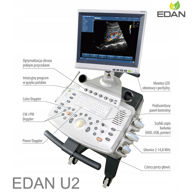
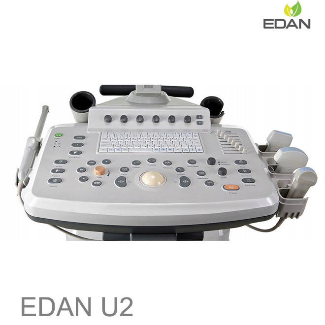
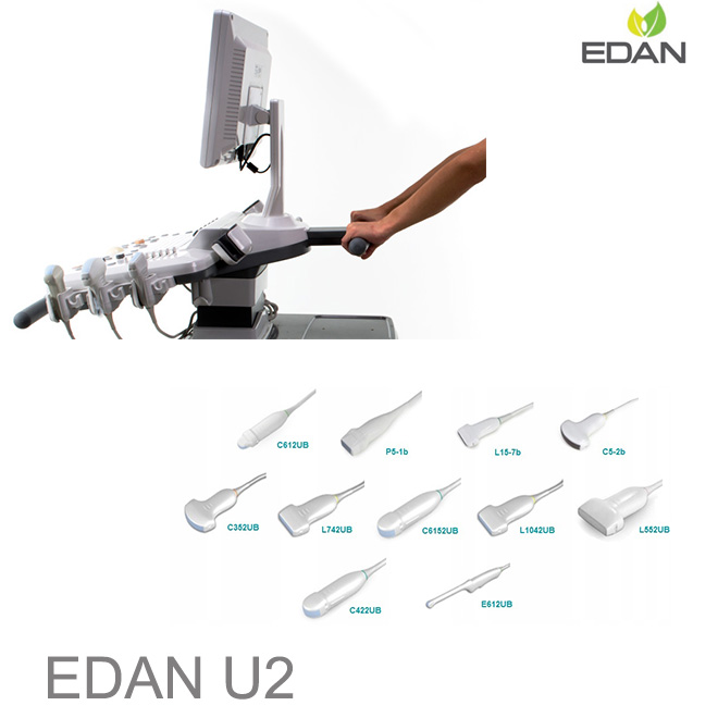
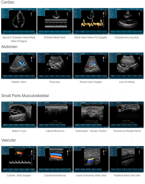
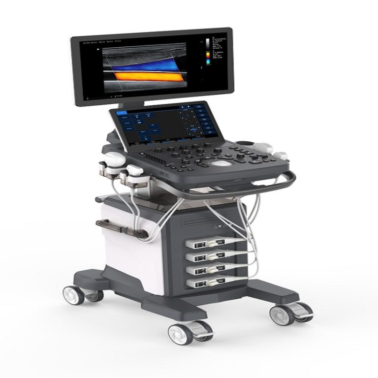
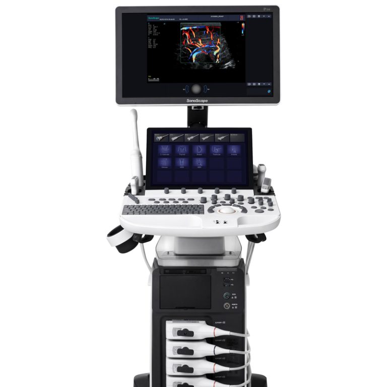
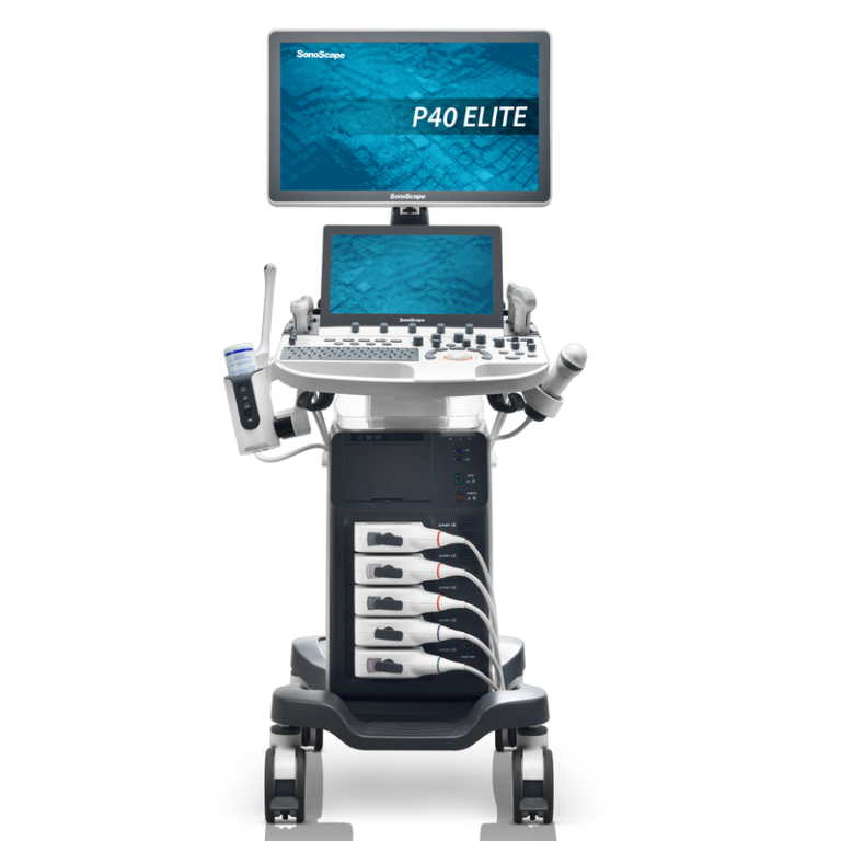
Imágenes armónicas con inversión de fase Reduce el ruido y el desorden para una calidad de imagen óptima Composición espacial Múltiples líneas de visión mejoran la resolución de contraste Tecnología multihaz Alta velocidad de fotogramas en todos los modos, incluidos color y Doppler Flujo de trabajo fácil de usar El panel de control intuitivo reduce la curva de aprendizaje Smart PreSet: ajuste rápidamente múltiples configuraciones para adaptarse a su tipo de paciente y preferencias de imagen Tecnología de transductor multifrecuencia Múltiples frecuencias 2-D, armónicas y de color aumentan la utilidad del transductor Aplicaciones compatibles Abdomen, obstetricia, ginecología, endovaginal, partes pequeñas, muculoesquelético, vascular, urología, cardiología, pediatría Diseño compacto de carro móvil, tiempo de arranque excepcionalmente rápido Batería de litio incorporada Monitor LCD de alta resolución de 15″ Múltiples puertos periféricos Tecnología de reducción de moteado Las imágenes con reducción de moteado utilizan procesamiento de imágenes en tiempo real para mejorar la visualización de la anatomía y la patología al reducir el ruido de moteado. La tecnología de imágenes con reducción de ruido de moteado de Edan utiliza un algoritmo avanzado de filtrado anisotrópico multiescala. Esta tecnología de imágenes es excelente para separar las regiones de ruido de la Imagen diagnóstica que permite realizar un filtrado complejo de forma diferente según el ruido o la información anatómica real, lo que produce una imagen mejorada. Imágenes armónicas con inversión de fase. Las señales armónicas se producen a medida que las ondas ultrasónicas se propagan por el cuerpo. Dado que estas señales se producen en el cuerpo, no se ven afectadas por la grasa que induce artefactos cerca de la superficie de la piel. En consecuencia, una imagen formada utilizando únicamente la señal armónica tendrá menos interferencias y puede ser más diagnóstica. Con armónicos de inversión de fase, se transmiten pares de pulsos ultrasónicos con fases opuestas. Al sumar las señales recibidas de los pulsos invertidos, se cancelan los componentes fundamentales y solo permanece la señal armónica. Esto crea una imagen armónica pura con reducción de los artefactos de interferencia que degradan la imagen. Composición espacial. La composición espacial combina varias imágenes de componentes diferentes para mostrar una imagen de mejor calidad. Además de la imagen formada mediante la transmisión de ondas ultrasónicas directamente desde el transductor, se forman imágenes con ondas dirigidas electrónicamente en un ángulo que se aleja del transductor. Se pueden utilizar diferentes grados de dirección para producir múltiples imágenes diferentes. El moteado en estas imágenes de componentes. Será diferente debido a la dirección electrónica, pero la variación macroscópica, como la variación de brillo causada por una lesión hepática, se compartirá entre las imágenes que lo componen. Al combinar las imágenes, se reduce el ruido de moteado y se mejora el contraste. Tecnología multihaz: Los sistemas de ultrasonido utilizan haces enfocados electrónicamente para producir una alta resolución espacial. Las señales del transductor se retardan y se suman en el sistema para producir un haz de ultrasonido enfocado. Las mismas señales se pueden sumar con diferentes retardos para producir múltiples haces. De esta manera, se puede mejorar la resolución espacial sin reducir la velocidad de fotogramas, o bien, se puede mejorar la velocidad de fotogramas sin reducir la resolución. Como se muestra a continuación, con la formación de haz dual, se forman los mismos haces de recepción con la mitad de los haces de transmisión, duplicando así la velocidad de fotogramas. Matriz convexa C352UB: Aplicaciones: Obstetricia/Ginecología, Abdomen, Pediatría, Urología. Frecuencias: 2,5/3,5/4,5/H5,0/H5,4 MHz. Matriz lineal L742UB: Aplicaciones: Piezas pequeñas, Vascular. Musculoesquelético, Superficial Frecuencias: 8/9,5/11,0/H13,0/H13,4 MHz Matriz microconvexa C6152UB Aplicaciones: Obstetricia/Ginecología, Abdomen, Pediatría, Urología Frecuencias: 5,5/6,5/7,5/H9,0/H9,4 MHz Matriz lineal L1042UB Aplicaciones: Partes pequeñas, Vascular, Musculoesquelético, Superficial Frecuencias: 8,0/9,5/11,0/H13,0/H13,4 MHz, Matriz microconvexa C612UB Aplicaciones: Obstetricia/Ginecología, Abdomen, Pediatría, Urología Frecuencias: 5,5/6,5/7,5/H9,0/H9,4 MHz Matriz lineal L552UB Aplicaciones: Partes pequeñas, Vascular, Musculoesquelético, Superficial, Pediatría Frecuencias: 4.5/ 5.5/ 6.5/ H5.6/ H6.0 MHz Matriz microconvexa C422UB Aplicaciones: Abdomen, Cardiología Frecuencias: 3.0/ 4.0/ 5.0/ H5.0/ H5.4 MHz Endovaginal E612UB Aplicaciones: Obstetricia, Ginecología Frecuencias: 5.5/ 6.5/ 7.5/ H9.0/ H9.4 MHz