
-
 Retiro en Casa Matriz
-
 Envío a Domicilio
-
 Certificado de Calidad
-
 Empaque y Traslado Seguro
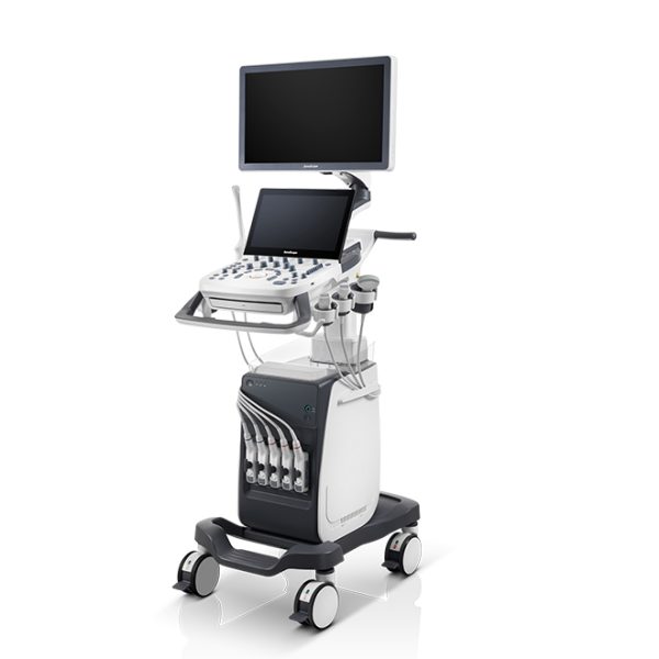
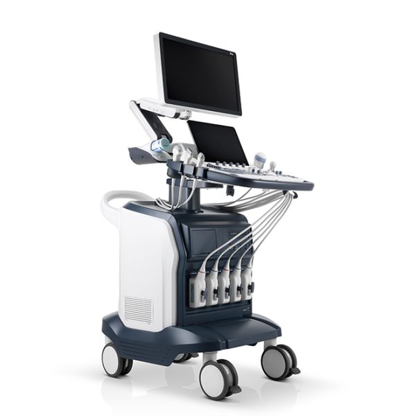
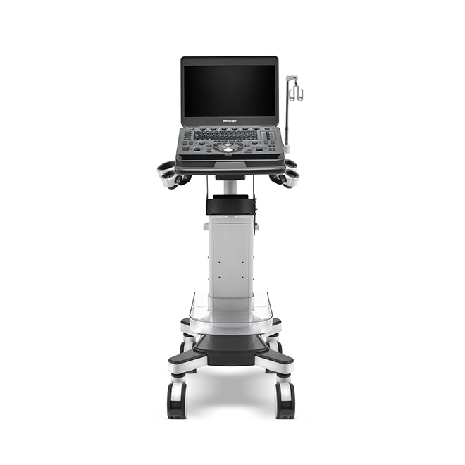
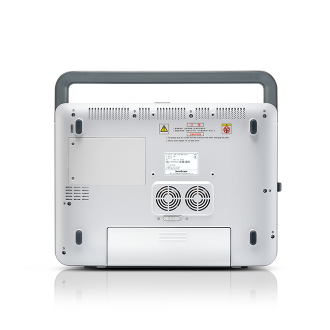
Dispositivo portátil de ultrasonido Doppler color veterinario Sonoscape X3V
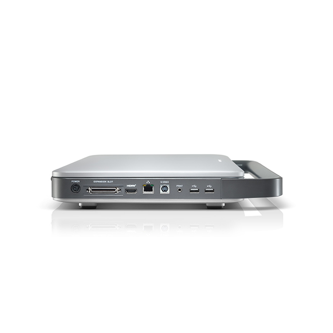
Unidad principal X3V
Monitor LED color de alta resolución de 15.6″
Un conector para transductor
USB 2.0/Disco duro de 500 G
Batería integrada
Adaptador de corriente
Dispositivo portátil de ultrasonido Doppler color veterinario Sonoscape X3V
1. Especificaciones generales El sistema adopta tecnologías avanzadas de Doppler ultrasónico, que incluyen el formador de haz de banda súper ancha digital completo, el enfoque dinámico digital, la apertura variable y Rastreo dinámico, rango dinámico de banda ancha y procesamiento paralelo multihaz, etc. Los usuarios pueden operar el sistema con un mínimo de capacitación u orientación. Este sistema ha sido diseñado para cumplir con las normas y regulaciones internacionales aplicables, lo que garantiza la seguridad y disponibilidad de este producto. Este sistema se basa en tecnología informática y el sistema operativo Linux, lo que lo hace más flexible y estable. El mantenimiento y la actualización de funciones del sistema se pueden realizar mediante actualizaciones de software, lo que aumenta el valor del producto y mantiene el avance tecnológico.
2. Especificaciones físicas
Dimensiones: 378 mm (ancho) × 61 mm (alto) × 340 mm (profundidad)
Peso: aprox. 4,5 kg (máximo, con batería) aprox. 4,1 kg (máximo, sin batería) Monitor: Pantalla ancha LCD a color de 15,6» y alta resolución, antiparpadeo y con rotación vertical y horizontal. Puerto para sonda: uno, ampliable a tres. 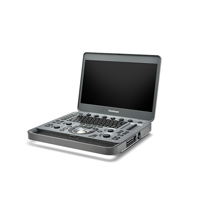 Ecografía Doppler Color Veterinaria Portátil Sonoscape X3V. 3. Tecnologías Avanzadas: Tecnología de interfaz digital. Tecnología de procesamiento multihaz. Imágenes compuestas espaciales. Tecnología de procesamiento de imágenes μScan. Imágenes armonizadas de tejidos. Imágenes armónicas invertidas Ícono de diagnóstico gráfico
 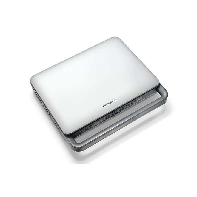 4. Configuraciones estándar Función μScan Frecuencia ajustable de 5 bandas en modo B Imágenes específicas de tejido (TSI) Modo THI Modo PHI Haces cuádruples Rotación de imagen Imágenes compuestas Imágenes trapezoidales (matriz lineal) Modo CFM Modo PDI Modo DPDI Modo CFMM Modo PW Modo CW Modo simultáneo B+PW Biopsia Optimización automática B/M Paquete de medición básica Paquete de medición de cardiología Paquete de medición de urología Paquete de medición de abdomen Paquete de medición vascular Paquete de medición de ginecología Paquete de medición de reproducción veterinaria Paquete de medición de tendones Índice TEI Trazado automático PW
Almacenamiento DICOM
Envío DICOM
5. Configuraciones opcionales
Mascota (departamento)
Carreras de caballos (departamento)
Ganadería (departamento)
Laboratorio (departamento)