
-
 Retiro en Casa Matriz
-
 Envío a Domicilio
-
 Certificado de Calidad
-
 Empaque y Traslado Seguro
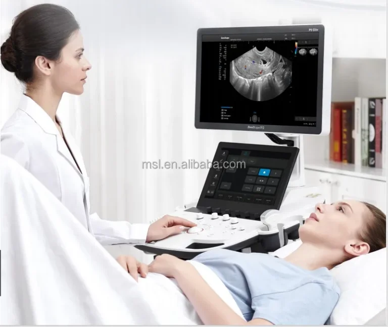
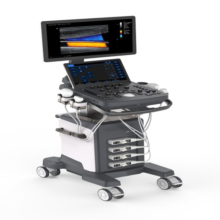
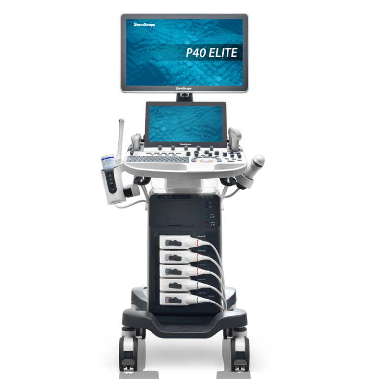
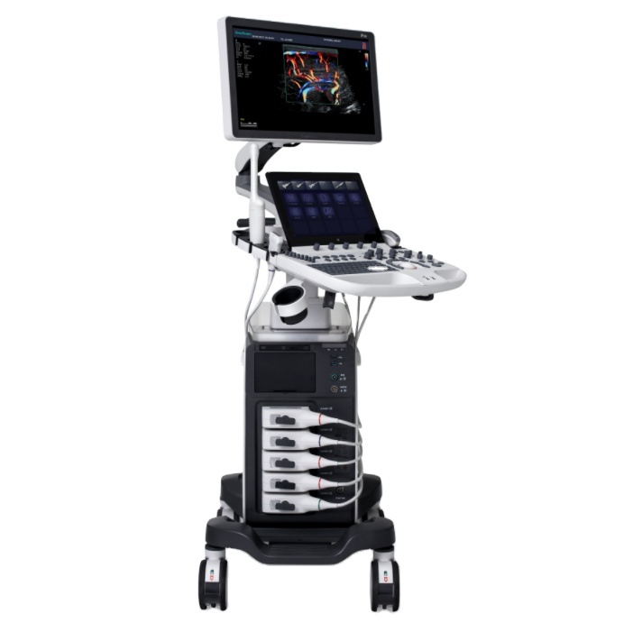
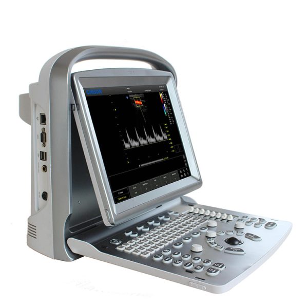
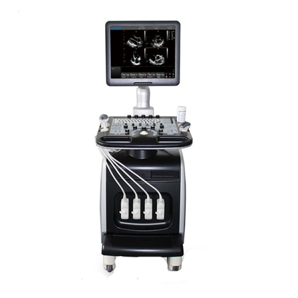
Ultrasonido 4D Doppler Obstétrico: Ultrasonido abdominal Chison i3 Ultrasonido 4D Doppler Obstétrico Chison i3 MODOS DE IMAGEN * B, 2B, 4B, B/M, M * CFM * Modo PW * Doppler de potencia/PD direccional * Trapezoidal * 4D en tiempo real (opcional) * Croma B/PW * CW (opcional) 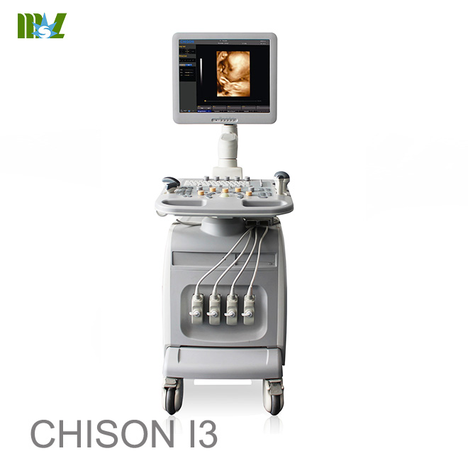 Ultrasonido 4D Doppler Obstétrico Chison i3 SONDAS * Sonda convexa * Sonda lineal * Sonda transvaginal * Sonda Phased Array * Sonda microconvexa * Sonda de volumen 4D Ultrasonido 4D Doppler Obstétrico Chison i3 PROCESAMIENTO DE IMÁGENES TECNOLOGÍA * AIO (movimiento dinámico de ajuste automático) * Imágenes trapezoidales * SRA (reducción de ruido de moteado) * THI * Tecnología compuesta * i-lmage: software de optimización de imágenes ultrasonido 4d doppler obstetrico chison i3 PAQUETES DE MEDICIÓN E INFORMES * OB&GYN * Vascular * Urología * Partes pequeñas * Cardíaco ultrasonido 4d doppler obstetrico chison i3 APLICACIONES CLÍNICAS AMPLIAS * Abdominal * * Vascular * Vascular periférico * Partes pequeñas * Próstata * Mama * Superficial * Musculoesquelético * Ginecológico y fertilidad * Imágenes generales pediátricas * Imágenes quirúrgicas * Obstétricas * Imágenes intervencionistas * Imágenes epicárdicas * Eco fetal ultrasonido 4d doppler obstetrico chison i3 CONFIGURACIÓN ESTÁNDAR Unidad principal, LCD de 19″, 4 conectores de sonda, Disco duro, DVD-RW, 6 memorias flash USB ultrasonido 4d doppler obstetrico chison i3 OPCIONES * Sonda convexa * Sonda lineal * Sonda lineal (60 mm) * Sonda transvaginal * Sonda transvaginal * Sonda de matriz en fase * Sonda microconvexa * Sonda microconvexa (cardíaca adulta) * Microconvexa * CW * ECG * Paquete 4D: incluye sonda de volumen 4D de 2,5 MHz a 5,3 MHz, software 4D y módulo de hardware 4D * Impresora de video (SONY UP-X898MD), impresora para PC (HP Pro 111P1102W y HP Pro 200 M251n y Canon selphy cp910) * DICOM 3.0 * i-lmage: software de optimización de imágenes * Kit de biopsia: para sonda convexa, lineal y de TV * Pedal * Superaguja * Dirección 2D * Cuádruple * IMT automático * Elastografía Nos reservamos el derecho a realizar cambios en este documento sin previo aviso Hoja de datos del sistema i3 Color Doppler V3.2 Nos reservamos el derecho a realizar cambios en este documento Información general Dimensiones y peso Dimensiones de la unidad principal (aprox.): 630 mm (ancho) × 1020 mm (profundidad) × 1365 mm (alto) Peso neto de la unidad principal (aprox.): 125 kg (sin sonda incluida) Alimentación eléctrica Voltaje de la fuente de alimentación: Adaptable automáticamente para CA 100-240 V Frecuencia de la fuente de alimentación: 50-60 Hz Consumo de energía: 600 VA Panel de operación Panel de control Teclado alfanumérico 8 diapositivas TGC Teclas retroiluminadas interactivas LCD a color de alta resolución – Dimensión diagonal: 19 pulgadas – Resolución: 1280 X 1024 – Ajuste de brillo Altavoz integrado – Volumen ajustable Descripción general del sistema Aplicaciones Abdomen Ginecología Obstetricia Urología Piezas pequeñas Nos reservamos el derecho de realizar cambios en este documento sin previo aviso Pediatría Vascular Musculoesquelético Cardíaco Método de escaneo Convexo electrónico Lineal electrónico Microconvexo electrónico Matriz en fase electrónica Convexo de volumen Tipos de transductor Sonda convexa de 3,5 MHz, Sonda lineal D3C60L de 7,5 MHz, sonda transvaginal D7L40L de 6,0 MHz, sonda microconvexa D6C12L de 3,0 MHz Sonda microconvexa D3C20L de 6,0 MHz Sonda de matriz en fase D6C15L de 3,0 MHz Sonda de volumen 4D D3P64L de 4,5 MHz V4C40L Modos de imagen Modo B Modo M PW (Doppler de onda de pulso) CW (opcional) CFM (Mapeo de flujo de color) CPA (Doppler de potencia) DPD (Doppler de potencia direccional) 4D (opcional) ECG (opcional) Imágenes trapezoidales (solo para sonda lineal) Modo de visualización Nos reservamos el derecho de realizar cambios en este documento sin previo aviso Pantalla cuádruple/dual (para B, CFM, CPA) Modo dúplex: B+CFM, B+PW, B+CPA, B+DPD, B/M Anotación en pantalla Nombre del hospital Fecha/hora Nombre e ID del paciente Estado del sistema (tiempo real o congelado) Barra de grises/color Guía de cine Dirección de escaneo Ventana de resumen de medición Ventana de resultados de medición Tipo de sonda Frecuencia Nombre de la aplicación Indicación de menú Indicación de funciones del trackball Parámetros de imagen mostrados en la pantalla Configuración estándar Pantalla LCD de 19 pulgadas de alta resolución 4 puertos de sonda activos Doppler de onda de pulso Imágenes de flujo Doppler color Imágenes de flujo Doppler de potencia Imágenes de flujo Doppler de potencia direccional ≥250 G de disco duro integrado Puertos USB: 6 (2 en el panel de control, 4 en el panel trasero) Puerto Ethernet Puerto de salida de S-video Puerto VGA Paquete de medición general Paquete de medición clínica Nos reservamos el derecho de realizar cambios en este documento sin previo aviso Pantalla en varios idiomas EasyView: sistema de archivo de imágenes Sistema de gestión de información del paciente Sistema de informes de edificios AIO (Optimización automática de imágenes) Algoritmo de reducción de moteado de zoom inteligente (SRA) Paquete de software i-Image TM Opciones de software Paquete de software DICOM 3.0 4D Opción de hardware de ECG CW Sonda convexa de 3,5 MHz, D3C60L Sonda lineal de 7,5 MHz, Sonda transvaginal D7L40L de 6,0 MHz, sonda microconvexa D6C12L de 3,0 MHz Sonda microconvexa D3C20L de 6,0 MHz Sonda de matriz en fase D6C15L de 3,0 MHz Sonda de volumen 4D D3P64L de 4,5 MHz, módulo V4C40L y 4D Pedal Cable de ECG Periféricos Impresora de video: SONY UP897MD Impresora para PC: – HP Laser Jet 1020 – HP Laser Jet CP2055d Nos reservamos el derecho a realizar cambios en este documento sin previo aviso Procesamiento y presentación de imágenes Modo B Potencia acústica Ganancia TGC Profundidad Frecuencia Velocidad de fotogramas Número de enfoque Posición de enfoque Ancho de escaneo Densidad de línea Persistencia dinámica Rechazo de ruido Suavizado Mejora de bordes i-Image TM SRA Mapa 2D compuesto Croma Gamma Brillo de pantalla Rotación de imagen Zoom Modo M Mapa de color Velocidad de barrido Diseño Nos reservamos el derecho a realizar cambios en este documento sin previo aviso Modo de color Ganancia Velocidad de fotogramas Dirección PRF Filtro de pared Mapa de color Flujo Color Invertir Densidad Persistencia Línea base Modo de color: Velocidad, Varianza Escala de efecto de sangre Modo CPA/DPD Ganancia Velocidad de fotogramas Dirección PRF Filtro de pared Mapa de color Flujo Densidad Persistencia Pared Thre. Modo PW Ganancia Escala PRF Invertir Filtro de pared Nos reservamos el derecho a realizar cambios en este documento sin previo aviso Audio Velocidad Línea base DA SV Mapa de color Mapa 2D Modo CW Ganancia Escala PRF Invertir Filtro de pared Audio Mapa de color Velocidad Línea base Mapa 2D Espectro dinámico Mejorar DA Almacenamiento Disco duro integrado ≥250 GB Controlador de DVD R/W Puertos USB Formato de almacenamiento de imágenes fijas: IMAG Formato de exportación de imágenes fijas: BMP, JPG, DCM, PNG, TIFF Formato de almacenamiento de bucles de cine: CINE Formato de exportación de bucles de cine: AVI Configuración de almacenamiento rápido: 3 s, 5 s, 10 s, tiempo personalizado, manual Nos reservamos el derecho a realizar cambios en este documento sin previo aviso EasyView Revisión de imágenes Diseño: 1×1,2×2 Gestión de imágenes – Eliminar imagen seleccionada – Exportar imagen seleccionada – Enviar imagen seleccionada a demostración – Imprimir imagen seleccionada con impresora de PC – Imprimir imagen seleccionada con impresora DICOM – Enviar imagen seleccionada con DICOM – Seleccionar todo – No seleccionar ninguno Revisión de examen BuscarExamen Revisión de examen: vista del paciente, vista del estudio Gestión de exámenes – Eliminar examen seleccionado – Exportar examen seleccionado – Hacer copia de seguridad del examen seleccionado – Recuperar desde la copia de seguridad del examen – Seleccionar todo – Expandir todo – Contraer todo – Editar examen seleccionado – Revisar examen seleccionado – Continuar examen seleccionado Medición y cálculo Paquete de medición general – Paquetes de software para diversos usos clínicos específicos – Métodos de análisis integrales – Informes de análisis clínicos Nos reservamos el derecho de realizar cambios en este documento sin previo aviso Paquete de medición general Modo B Medición normal Distancia Longitud_Área(Elipse) Longitud_Área(Trazo) Volumen(1 Distancia) Volumen(2 Distancia) Volumen(3 Distancia) Volumen(1 Elipse) Volumen (2 Elipse) Volumen (1 Distancia 1 Elipse) Relación Ángulo Modo M Medición normal Distancia media Tiempo medio Velocidad Frecuencia cardíaca Modo PW Medición normal Velocidad Distancia Pico Trazo automático Trazo manual FC Flujo Volumen StD% StA% Área Análisis clínico Paquetes OB Medición OB-B Distancia Biometría fetal: GS, CRL, YS, BPD, OFD, HC_Ellipse, APD, TAD, AC(Elipse), Nos reservamos el derecho de realizar cambios en este documento sin previo aviso FTA, FL, SL, APTD, TTD, ThC Huesos largos fetales: Húmero, CÚBITO, Tibia, RAD, FIB, CLAV Fetal Cráneo: CER, CM, NF, NT, OOD, IOD, NB, Lvent, HW OBOtros: LtKid, RtKid, LtRenalAP, RtRenalAP, LVWrHEM, MAD AFI: AFI_1, AFI_2, AFI_3, AFI_4 FBP: AF Ductus venoso: StA%, StD%, área del vaso, Dis del vaso StA%: A Out, A En StD%:D Out,D En CX_L Aorta:StA%,StD%,Veslumen_D,Veslntimal_D,VesOutside_D,Veslntimal_A, Veslumen_A StA%:A Out,A In StD%:D Out,D In Aorta descendente:StA%,StD%,Veslumen_D,Veslntimal_D,VesOutside_D, Veslntimal_A,Veslumen_A StA%:A Out,A In StD%:D Out,D In MCA:StA%,StD%, Veslumen_D, Veslntimal_D, VesOutside_D, Veslntimal_A, Veslumen_A StA%: A Out, A In StD%: D Out, D In UmbA:StA%,StD%,Veslumen_D,Veslntimal_D,VesOutside_D,Veslntimal_A, Veslumen_A StA%:A Out,A In StD%:D Out,D In Arteria uterina:Arteria uterina (Rt), Arteria uterina (Lt) Arteria uterina (Rt): StA%,StD%, Veslumen_D, Veslntimal_D, VesOutside_D, Veslntimal_A, Veslumen_A Arteria uterina (Lt): StA%, StD%, Veslumen_D, Veslntimal_D, VesOutside_D, Veslntimal_A, Veslumen_A StA%: A Out, A In StD%: D Out, D In Arteria pulmonar: StA%, StD%, Veslumen_D, Veslntimal_D, VesOutside_D, Veslntimal_A, Veslumen_A StA%: A Out, A In Nos reservamos el derecho de realizar cambios a este documento sin previo aviso StD%: D Out, D In Selección fetal Medida OB–D Umb A Aorta Aorta descendente Arteria uterina (Izq.) Arteria uterina (Der.) Arteria pulmonar ACM Medida FCF Medida OB–M Distancia Mtiempo Velocidad Frecuencia cardíaca GYN Medida GYN–B Distancia UT: UT_L, CX_L, UT_W, UT_H Vol. cérvix: Longitud, Altura, Ancho ENDO Volumen OV derecho: Longitud, Altura, Ancho Volumen OV izquierdo: Longitud, Altura, Ancho FO_D derecho: Longitud, Ancho FO_D izquierdo: Longitud, Ancho Arteria uterina: Arteria uterina (D), Arteria uterina (I), Arteria uterina (D): % EstA, % EstD, Área vascular, Vaso Dis Arteria uterina (I): % EstA, % EstD, Vaso Área, Vaso Dis StA%: A Salida, A Entrada StD%: D Salida, D Entrada GYN –Dmeasure Umb A: Umb A (D), Umb A (Izq.) MCA: MCA (D), MCA (Izq.) Dt Uterina A Izq. Uterina A FetalAO: Fetal AO (D), FetalAO (Izq.) FHR GYN –M medida Nos reservamos el derecho de hacer cambios a este documento sin previo aviso Mdistancia Mtiempo Velocidad Frecuencia cardíaca Pediatría CADERA URO Distancia Vol. residual Vol. próstata Riñón izquierdo Riñón derecho Vol. zona T Vol. vejiga StA% StD% Área del vaso Vaso Dis Cardíaco Medida Cardíaca-B Distancia Plano único: Biplano Volumen de bala Modi_Simpson Teichholz Cube LV/RV AO/LV LVOT MV AV Cardiac-D measure Nos reservamos el derecho de realizar cambios a este documento sin previo aviso LVOT AV MV TV PV Pul.Vein HR Cardiac-M measure Distance Heart_Rate Eyection_Time LV LVSHORT AV AVSHORT MV AV AO/LV LVOT TV PulV Vessel Prox CCA Mid CCA Distal CCA Prox ICA Mid ICA Distal ECA Vertebral A INT IIL EXT IL ILIAC CFA ProFun LTCIR SFA Nos reservamos el derecho de realizar cambios a este documento sin previo aviso Pop A ATA PTA PERON DRPED Abdomen CBD GB Wall Liver Length Bladder Bladder Prox Aorta Mid Aorta Distal Aorta Esplenomegalia Renal Vol. Iliac Carótida Subclavia A Prox CCA Mid CCA Distal Bulb Prox ICA Mid ICA Distal ECA Vertebral A Medición General Flujo Volumen Piezas pequeñas Relación de medición general Nos reservamos el derecho de realizar cambios en este documento sin previo aviso Configuración del sistema de ángulos Al usar la configuración del sistema, los usuarios pueden Personalizar la información del hospital Personalizar el idioma Personalizar el tiempo de almacenamiento rápido Personalizar el mapa de colores Asignar funciones al botón «IMPRIMIR» en el panel de control y el pedal Personalizar la biblioteca de comentarios Personalizar el informe Funciones definidas por el usuario Mediante la función definida por el usuario, los usuarios pueden personalizar el ajuste preestablecido definido por el usuario, que incluye: – Nombre de las aplicaciones, Nombre de los ajustes preestablecidos, Nombre definido por el usuario – Tipo de examen de las aplicaciones – Parámetros de imágenes Interfaz de pantalla en varios idiomas Inglés Chino Francés Español Ruso Polaco Portugués Sistema operativo Windows XP Embedded Nos reservamos el derecho de realizar cambios en este documento sin previo aviso Transductores Selección del transductor Nombre Tipo de matriz Frecuencia central D3C60L Convexo 3,5 MHz D7L40L Lineal 7,5 MHz D6C12L Microconvexo 6,0 MHz V4C40L Convexo 4,5 MHz D3C20L Microconvexo 3,0 MHz D6C15L Microconvexo 6,0 MHz D3P64L Phased Array 3.0MHz Entradas y salidas S-video: 1 Salida de video: 1 VGA: 2 Puerto USB: 6 Ethernet: 1 Control remoto: 1 Puerto de pedal: 2 Entrada de alimentación del sistema: 1 Polo de tierra: 1 Botón de encendido: 1 Condiciones de funcionamiento Temperatura ambiente: 10 °C a 40 °C Humedad relativa: 30 % a 75 % (sin condensación) Presión atmosférica: 700 hPa a 1060 hPa Condiciones de almacenamiento Temperatura ambiente: -5 °C a 40 °C Humedad relativa: ≤ 80 % (sin condensación) Nos reservamos el derecho de realizar cambios en este documento sin previo aviso Presión atmosférica: 700 hPa a 1060 hPa Estándares de calidad ISO 10993 Evaluación biológica de dispositivos médicos IEC 60601-1 Equipos médicos eléctricos IEC 60601-1-1 Equipos médicos eléctricos IEC 60601-1-2 Compatibilidad electromagnética IEC 60601-1-4 Sistemas médicos programables IEC 60601-2-37 Requisitos particulares para la seguridad de equipos de diagnóstico y monitorización médica por ultrasonidos. Es posible que no todas las características o especificaciones descritas en este documento estén disponibles en todas las sondas o modos. CHISON Medical Imaging Co., Ltd. se reserva el derecho de modificar las especificaciones y características que se muestran aquí, o de discontinuar el producto en cualquier momento sin previo aviso ni obligación. Para obtener la información más actualizada, contacte con un representante de CHISON. Ultrasonido Sonoscape en oferta | Lista de precios de ultrasonido Chison | Precio del SonoScape X5 | Precio del SonoScape S9 | Precio del Sonoscape S8 | Precio del Sonoscape S8 EXP | Sonoscape A5 | Sonoscape S2 | Sonoscape A6 | Sonoscape S6 | Sonoscape A6VET | Sonoscape S40 | Sonoscape S50 | Sonoscape S22 | Sonoscape S12 | Sonoscape S30 | Sonoscape S11 | SonoScape X3 Precio del Sonoscape S20, precio del Chison Q5, precio del Chison Q9, precio del Chison Ebit 50, precio del Chison Ebit 60, precio del Chison I3, precio del Chison I8, precio del Chison EC01, precio del Chison EC03exp, precio del Chison EC05, precio del Chison EC06. Imagen del equipo B.S.D. Certificado B.S.D. B.S.D. Medical colabora con DHL, FEDEX, UPS, EMS, TNT, etc. Empresa de transporte internacional que garantiza la llegada segura y rápida de sus productos a su destino.